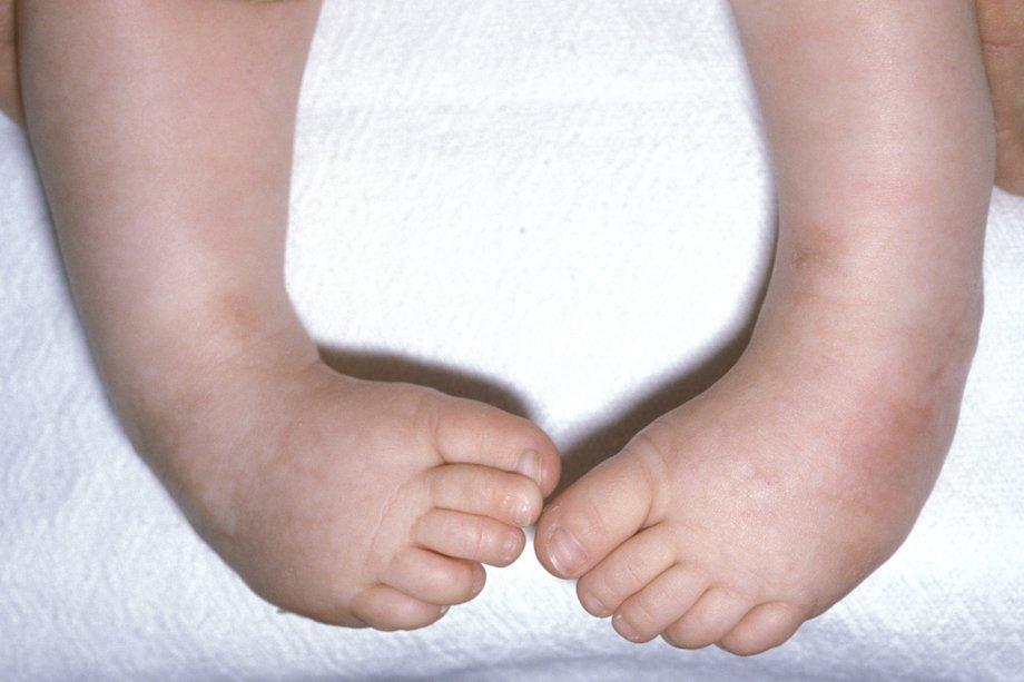
CTEV is a complex congenital deformity of foot in which a baby is born with a foot (or both feet) twisted out of its normal position. The condition affects bones, muscles, tendons, and blood vessels.
Components of the Deformity
The name “Talipes Equinovarus” describes the main components:
- Talipes: Involving the ankle (talus bone)
- Equinus: The foot points downward (like a horse’s hoof)
- Varus: The heel turns inward
So, in CTEV, the foot is:
- Plantarflexed (toes point down)
- Inverted (sole turns inward)
- Adducted (forefoot turns toward the midline)
Epidemiology
- Occurs in about 1 in 1,000 live births
- More common in males
- Bilateral in about 50% of cases
Causes
The exact cause of CTEV is often unknown, but it is considered multifactorial, involving a combination of genetic, environmental, and possibly mechanical factors. Here’s a detailed breakdown:
1. Genetic Factors
- Family history: CTEV is more likely if a parent or sibling had it.
- Polygenic inheritance: Multiple genes are thought to contribute.
- Studies show a higher concordance rate in identical twins, suggesting a strong genetic component.
2. Environmental and Maternal Factors
- Uterine constraint: Limited space in the womb may restrict fetal movement (e.g., in multiple pregnancies, oligohydramnios).
- Smoking during pregnancy: Increases the risk significantly.
- Drug use: Certain medications taken during pregnancy may play a role.
3. Neuromuscular Disorders (Secondary Clubfoot)
These are non-idiopathic causes, where CTEV is part of a broader condition:
- Spina bifida
- Cerebral palsy
- Arthrogryposis multiplex congenita
- Poliomyelitis (rare today)
Clinical features
CTEV is typically diagnosed at birth (or sometimes prenatally via ultrasound). The hallmark clinical features involve visible foot deformity and structural abnormalities.
Equinus
- The foot is plantarflexed (toes point downward).
- Heel is elevated and cannot touch the ground.
Varus
- The heel turns inward (inversion).
- Hindfoot is deviated medially.
Adduction of the Forefoot
- The front part of the foot is turned medially toward the midline.
Cavus Deformity
- High medial longitudinal arch due to contracture of intrinsic foot muscles.
Medial Rotation of the Tibia
- The leg may appear rotated, contributing to the overall twisted appearance.
Calf Muscle Atrophy
- Affected leg may have thinner and shorter calf muscles.
Smaller Foot Size
- The affected foot is smaller and shorter than normal.
Stiffness
- The deformity is rigid in untreated cases.
- Passive correction is limited.
Diagnosis
The diagnosis of CTEV is primarily clinical, based on visual inspection and physical examination. Early and accurate diagnosis is essential for prompt treatment to prevent long-term disability.
1. Clinical Diagnosis (Main Method)
At Birth:
- Diagnosis is usually evident immediately upon delivery.
- Inspection reveals the classic four deformities:
- Equinus (plantarflexed foot)
- Varus (heel tilted inward)
- Adduction (forefoot turned medially)
- Cavus (high medial arch)
Key Points:
- The deformity is rigid or semi-rigid.
- Cannot be fully corrected passively.
- Examine for calf muscle wasting and leg length discrepancy.
🔍 2. Antenatal (Prenatal) Diagnosis
- Ultrasound at 18–24 weeks gestation may detect CTEV.
- Seen as abnormal positioning of the feet.
- Helps plan for early orthopedic intervention.
- Prenatal diagnosis may also trigger genetic testing if other anomalies are found.
📊 3. Imaging (If Needed)
Not routinely required in typical cases, but used in special situations:
- X-rays:
- Useful in older children, recurrent, or atypical cases.
- Helps assess bony alignment, especially the talocalcaneal and talo-first metatarsal angles.
- Challenging in neonates due to unossified bones.
- MRI or CT:
- Rarely used
- Reserved for complex, syndromic, or surgical planning cases.
🧪 4. Assessment Tools (for Severity & Progress)
- Pirani Score (0–6):
- Evaluates severity based on six foot signs
- Higher score = more severe deformity
- Dimeglio Classification:
- Another grading scale to assess rigidity and reducibility
Physiotherapy management
Physiotherapy plays a crucial role in both conservative and post-surgical management of CTEV. It helps in correcting the deformity, maintaining flexibility, and preventing recurrence.
Goals of Physiotherapy
- Correct the foot deformity
- Improve range of motion (ROM)
- Strengthen foot and leg muscles
- Maintain functional alignment
- Prevent recurrence or complications
1. Early Conservative Management (Typically in First Weeks of Life)
a. Stretching and Manipulation
- Gentle, daily passive stretching of:
- Achilles tendon (for equinus)
- Medial foot structures (for adduction/varus)
- Hold stretches for 10–30 seconds, repeated multiple times a day.
b. Serial Casting (Part of Ponseti Method)
- Done by orthopedic specialists, but physiotherapists may assist or support.
- After manipulation, the foot is held in corrected position using plaster casts.
- Casts are changed weekly for 5–8 weeks.
- Often followed by a percutaneous Achilles tenotomy.
2. Post-Casting/Botox/Tenotomy Phase
a. Bracing (Maintenance Phase)
- Foot abduction brace (boots and bar) used:
- 23 hours/day for first 3 months
- Then night-time and naps for up to 4–5 years
- Physiotherapists educate and assist parents in brace application and compliance.
b. Range of Motion Exercises
- Gentle ROM exercises to maintain flexibility.
- Focus on:
- Dorsiflexion
- Eversion
- Forefoot abduction
c. Strengthening
- Encourage active movements as the child grows.
- Use play-based therapy to stimulate ankle and foot muscle activation.
3. Post-Surgical Physiotherapy (If Surgery Was Needed)
a. Pain and Swelling Management
- Ice therapy (if indicated)
- Elevation
b. Mobilization
- Gradual weight-bearing and gait training
- Stretching tight structures
- Scar tissue massage (to prevent adhesions)
c. Orthotic Support
- Advice on custom orthotics or shoes, if needed
- Monitor for pressure areas or improper fit
What does CTEV stand for, and what is its basic definition?
CTEV stands for Congenital Talipes Equinovarus. It is a complex congenital deformity where a baby is born with one or both feet twisted out of their normal position, affecting bones, muscles, tendons, and blood vessels.
What are the key clinical features of CTEV observed at birth?
The key clinical features include a plantarflexed foot (equinus), inward-turned heel (varus), medially turned forefoot (adduction), high medial arch (cavus), and possible calf muscle atrophy.
How is physiotherapy used in the management of CTEV?
Physiotherapy helps correct the deformity, maintain flexibility, and prevent recurrence through stretching, manipulation, ROM exercises, strengthening, and post-casting bracing support.

