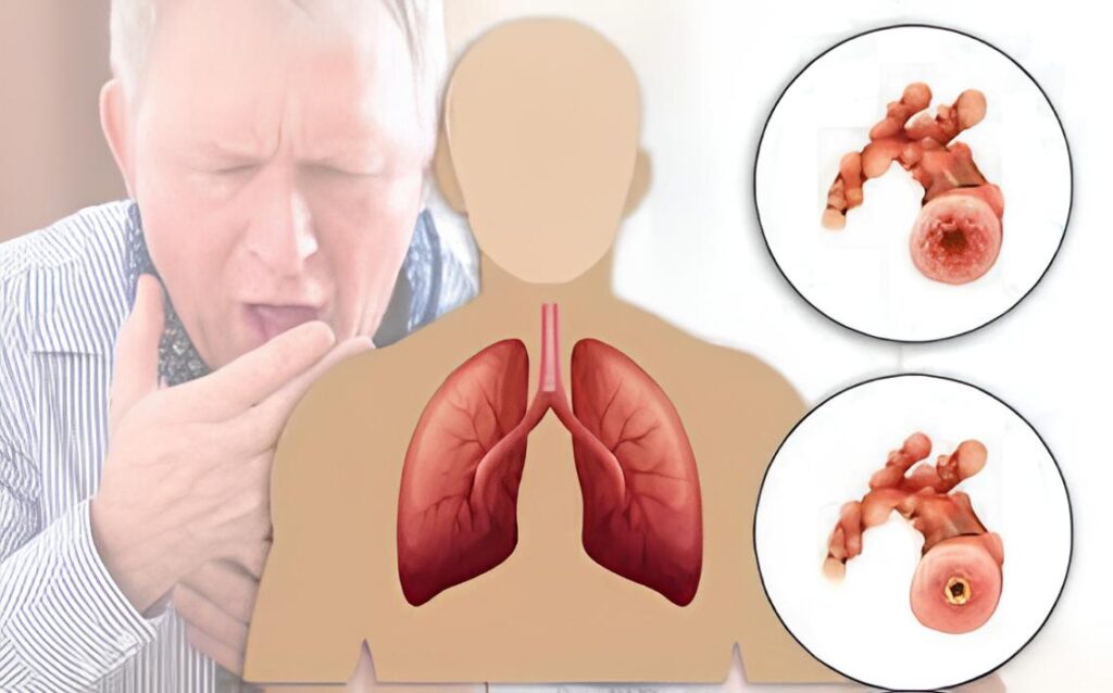
Chronic Obstructive Pulmonary Disease (COPD) is a group of progressive lung diseases that cause obstructed airflow from the lungs. The two most common conditions under COPD are:
- Chronic Bronchitis – Long-term inflammation of the bronchial tubes with excess mucus production.
- Emphysema – Gradual destruction of the alveoli (air sacs), leading to decreased oxygen exchange.
COPD worsens over time and can significantly limit quality of life. It is preventable and treatable, especially when diagnosed early.
Causes
- Cigarette smoking is the leading cause of COPD. Around 85–90% of COPD cases are
Risk Factors
- Long-term exposure to:
- Air pollution
- Dust and chemical fumes in the workplace
- Secondhand smoke
- Genetic factors (e.g., Alpha-1 Antitrypsin Deficiency – a rare inherited condition)
- Frequent respiratory infections in childhood
- Age – risk increases after age 40
- Asthma – especially if poorly controlled
Pathophysiology
COPD involves both airflow limitation and destruction of lung tissue:
- In chronic bronchitis, the airways become inflamed and narrowed. Mucus glands enlarge, producing excessive mucus.
- In emphysema, the walls between alveoli break down, reducing surface area for gas exchange. The lungs lose elasticity, making it harder to exhale.
These changes trap air in the lungs, reduce oxygen intake, and make breathing inefficient.
Symptoms
Symptoms develop slowly and worsen over time:
- Chronic cough (usually with phlegm)
- Shortness of breath (especially during exertion)
- Wheezing
- Chest tightness
- Frequent respiratory infections
- Fatigue
- Weight loss (in advanced stages)
- Cyanosis (bluish lips/fingernails – in severe disease)
Diagnosis
1. Medical History & Physical Exam
- Smoking history
- Environmental exposures
- Symptom history
2. Pulmonary Function Tests (PFTs)
- Spirometry is the gold standard:
- Measures FEV₁ (Forced Expiratory Volume in 1 second)
- FVC (Forced Vital Capacity)
- FEV₁/FVC ratio < 70% confirms airflow limitation
3. Imaging
- Chest X-ray – can show hyperinflation, flattened diaphragm
- CT scan – more detailed; can detect emphysema
4. Blood Tests
- Arterial blood gases – in advanced COPD to check oxygen and CO₂ levels
- Alpha-1 antitrypsin test – if under 45 or with family history
Stages of COPD (GOLD Criteria)
Based on FEV₁ value after bronchodilator:
| Stage | Severity | FEV₁ (% predicted) |
|---|---|---|
| GOLD 1 | Mild | ≥ 80% |
| GOLD 2 | Moderate | 50–79% |
| GOLD 3 | Severe | 30–49% |
| GOLD 4 | Very Severe | < 30% |
Symptoms and exacerbation risk also influence treatment decisions.
Physiotherapy management
Goals of Physiotherapy in COPD
- Improve ventilation and oxygenation
- Reduce dyspnea
- Mobilize and clear secretions
- Enhance exercise tolerance
- Promote independence in daily activities
- Educate patients on breathing strategies and energy conservation
🔑 Key Physiotherapy Interventions
1. Breathing Techniques
✅ Pursed-Lip Breathing
- Slows breathing and prevents airway collapse
- Reduces breathlessness
- How: Inhale through the nose → exhale slowly through pursed lips
✅ Diaphragmatic (Abdominal) Breathing
- Encourages efficient use of the diaphragm
- Reduces accessory muscle use
- How: Patient places one hand on the abdomen and one on the chest; focus on expanding the belly during inhalation
2. Airway Clearance Techniques (ACTs)
Important for those with chronic bronchitis or excessive mucus.
🔹 Active Cycle of Breathing Technique (ACBT)
- Combines breathing control, deep breathing, and huffing
- Helps loosen and expel mucus
🔹 Huff Coughing
- Gentle but forceful exhalation with an open mouth to mobilize mucus without straining
🔹 Postural Drainage
- Uses gravity to assist mucus drainage from specific lung segments
Percussion and Vibration
- Manual or mechanical techniques to loosen secretions (used cautiously)
Positive Expiratory Pressure (PEP) Devices
- Devices like Flutter or Acapella improve airway clearance
3. Pulmonary Rehabilitation
A structured multidisciplinary program typically lasting 6–12 weeks.
♂️ Components:
- Exercise training: Endurance (e.g., walking, cycling) and resistance training
- Education: COPD knowledge, inhaler technique, nutrition
- Psychosocial support: Anxiety/depression management
- Outcome monitoring: 6-minute walk test, dyspnea scales (e.g., MRC, Borg)
♀️ Exercise Benefits:
- Improves VO₂ max, reduces dyspnea
- Boosts muscle strength and cardiovascular fitness
Reduces hospital admissions
Q. What are the two most common conditions under COPD?
The two most common conditions are chronic bronchitis and emphysema.
Q: What is the gold standard test for diagnosing COPD?
Spirometry is the gold standard, measuring FEV₁, FVC, and the FEV₁/FVC ratio.
Q: Name one key breathing technique used in physiotherapy for COPD.
Pursed-lip breathing is a key technique that helps reduce breathlessness and prevent airway collapse.