Introduction
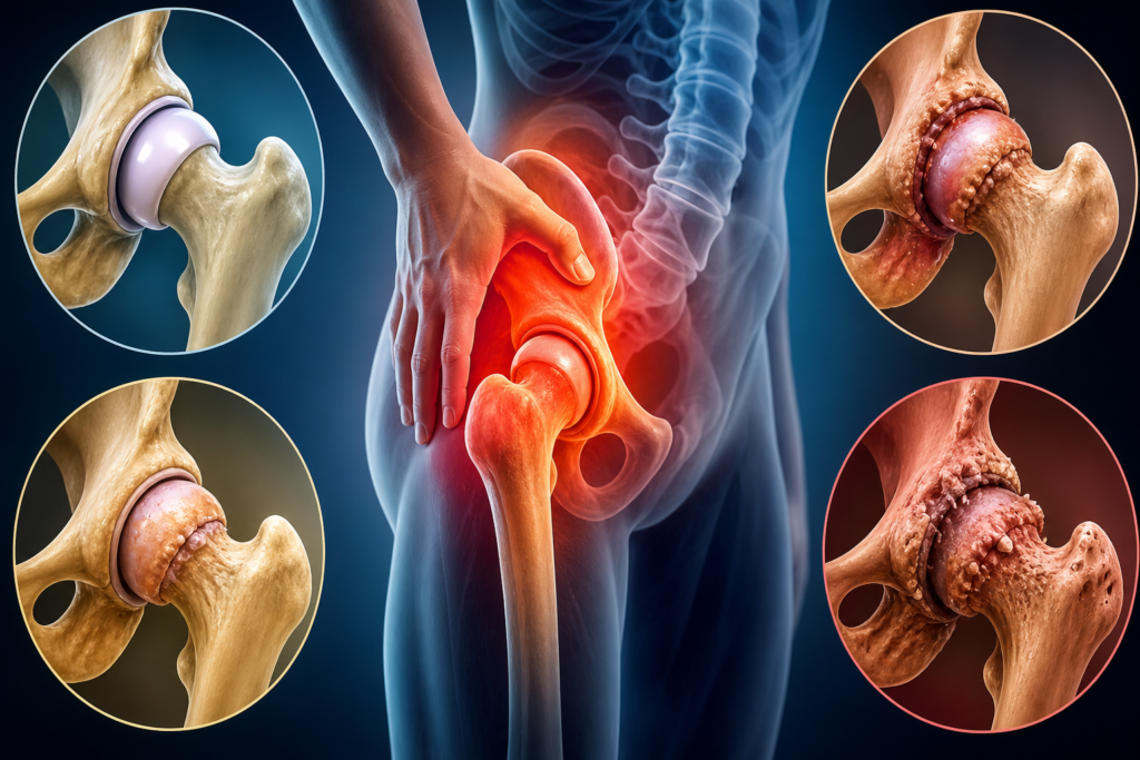
Tuberculosis of the Hip Joint constitutes 15 percent of all osteoarticular tuberculosis. It is always secondary. The initial focus of infection could be either in the: (i) acetabular roof, (ii) epiphysis, (iii) metaphyseal region, (iv) greater trochanter, (v) synovial membrane (rare), and (vi) trochanteric bursae
Pathogenesis
- Following injury, the vessels rupture and there is hemorrhage. The tubercle bacilli present in the circulation settle and proliferate in the blood clot so initiate.A tubercle follicle is formed and it consists of giant cells, lympho cytes, and endothelial cells . Small such tubercle follicles coalesce to form a larger follicle, which undergoes caseation at the center and fibrosis at the periphery.
- The caseation at the center of the shaft breaks down forming pus. It spreads towards the subperiosteal region, breaks the periosteum, and tracks along the lines of least resistance.
- It reaches the skin and forms the cold abscess (not warm). Later on, it breaches the skin forming the sinus.
Etiology
TB bacillus is may responsible for TB of the Hip Joint.
Route: Always secondary, may spread to the bone through.
Precipitating factors: General factors like anemia, debility, etc. help precipitate the infection.
Local factors: Like trauma, etc. localize the problem to the bone. Local trauma causes vascular stasis and intraosseous hemorrhage.
Clinical Features
- Tuberculosis of hip is common in the first three decades of life. The patient usually presents with painful limp and is the most common earliest symptom.
- He or she has an antalgic gait with a short stance phase.
- Pain is maximum towards the end of the day and there is a history of nocturnal cries.
- There is marked wasting of the thigh and gluteal muscles. There may be presence of sinuses and scars .
- Tenderness can be elicited by direct pressure in the femoral triangle or by bitrochanteric compression.
Deformities
- Flexion deformity: Flexion deformity in the initial stages of the disease, patient keeps the hip in flexion, as this is the position of ease and of maximum joint capacity.
- Adduction deformity: Soft tissue contractures permute the adduction position adapted by the patient due to the spasm of the adductor muscles following damage to the articular cartilage, to one of the fixed adduction deformities.
The adduction deformity can be revealed by squaring the pelvis. This is done by adducting the affected limb until both the anterosuperior iliac spines lie in the same straight line. The angle formed between the vertical and the adducted limb is the angle of fixed adduction deformity.
- Scoliosis deformity : Scoliosis deformity of the spine towards the affected side. The fixed abduction deformity can be revealed by abducting the affected limb until both the anterosuperior iliac spine lie in the same level.
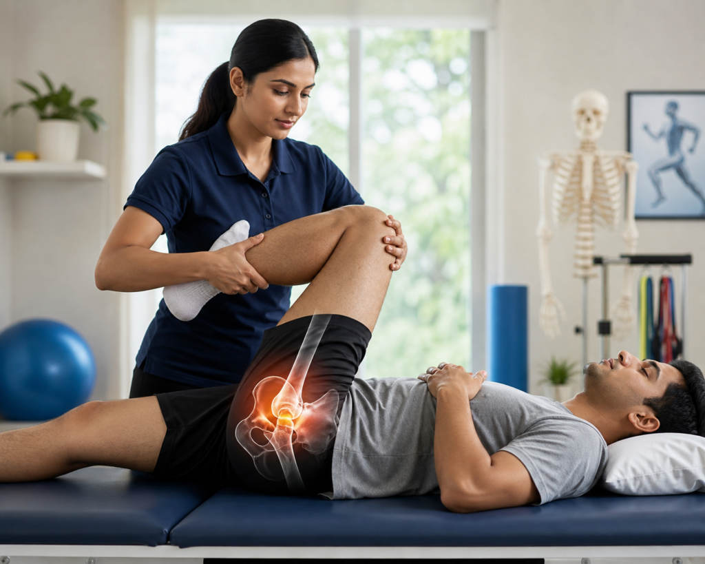
Physiotherapy Management
Physiotherapy plays a vital role in conjunction with antitubercular drug therapy and, if necessary, surgical intervention.
- Pain Management: Modalities like ice, heat, and gentle manipulation therapy.
- Range of Motion (ROM) Exercises: Gentle, progressive exercises to maintain and improve joint mobility, avoiding forced movements.
- Hydrotherapy: Buoyancy can aid in uncomplicated movement.
- Gait Training: Crutch or wheelchair training to minimize weight-bearing during the acute phase.
- Strengthening Exercises: Isometric exercises for hip adductors, abductors, extensors and flexors, to prevent muscle atrophy.
- Splinting/Bracing: To prevent or correct malformation and provide joint rest.
- Patient Education: Emphasizing adherence to medication and activity modification.
Dietary Management
A nutrient-rich diet plays an essential role in the recovery and rehabilitation of hip tuberculosis patients.
Proper nutrition enhances immunity, supports tissue healing, and helps the body respond better to anti-tubercular medications.
Key dietary guidelines:
- High-protein foods: Include eggs, lentils, lean meats, soy, and dairy to rebuild muscle mass and repair tissues.
- Vitamin-rich fruits: Citrus fruits, guava, and mud apple (sapodilla) provide vitamin C and antioxidants that aid healing and immune function.
- Iron and calcium intake: Green leafy vegetables, nuts, and seeds help counter anemia and strengthen bones.
- Adequate hydration: Promotes detoxification and better nutrient absorption.
- Avoid processed or oily foods: These may cause inflammation and interfere with medication absorption.
Conclusion
Tuberculosis of the hip joint is a debilitating condition requiring a high index of suspicion for early diagnosis. A multidisciplinary approach involving antitubercular chemotherapy, surgical intervention where indicated, and overarching physiotherapy management is paramount to achieve optimal functional outcomes, intercept deformities, and improve the quality of life for affected individuals.
1. What is the main goal of physiotherapy in tuberculosis of the hip joint?
Answer: The main goal is to reduce pain, maintain joint mobility, prevent deformities, and restore functional movement.
2. Which modalities are used for pain management in hip tuberculosis?
Answer:Ice, heat, and gentle manipulation therapy are used for pain relief.
3. Why are range of motion (ROM) exercises important?
Answer:A3. ROM exercises help maintain and improve joint mobility without causing strain.