Introduction
Avascular Necrosis Hip stands out due to its progressive charecter and potential for severe joint damage if left unaddressed. This article delves into the various facets of AVN of the hip.
Definition
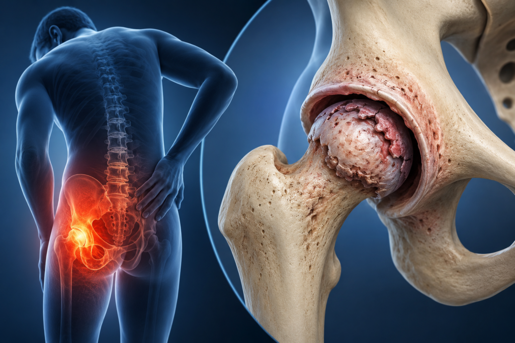
Avascular Necrosis Hip (AVN), also called osteonecrosis, is caused by disruption of blood supply to the femoral head.
Etiology
Avascular Necrosis Hip can be caused due to injury to the hip joint or due to other causes.
- Post-traumatic: Fracture of the femoral neck
- Dislocation of the hip
- Nontraumatic: Steroid-induced
Organ transplant
Alcoolism
Sickle cell disease
Systemic lupus erythematosus (SLE)
Postirradiation .
Clinical Features
In early stages, pain might be intermittent, but as the condition progresses, it becomes constant
- The patient presents with pain and limitation of movements at the affected joint with a limp.
- there is limitation of movements at the hip joint
- Internal rotation is painful.
Investigation
1. X-ray of the hip shows increased sclerosis, cyst formation and deformation (in the later stages of the disease) of the femoral head. The disease can be staged radiographically.
2. MRI is the gold standard in the diagnosis of AVN with a sensitivity and specificity of more than 98%.
Risk Factors
Key risk factors include high-dose or long-term corticosteroid therapy, hip trauma, heavy alcohol intake, certain blood disorders, Gaucher’s disease, and decompression sickness. Age and gender can also play a role, with men between 30 and 50 years being more commonly affected.
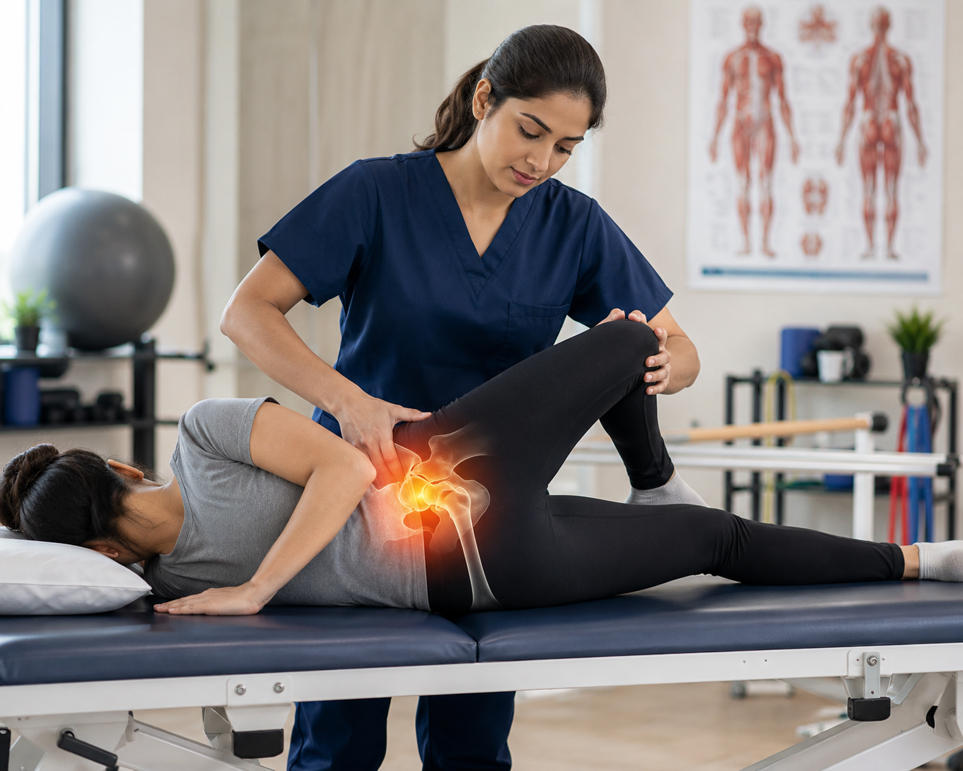
Physiotherapy Management
The approach of physiotherapy depends upon the surgical procedure adopted.
Non–weight-bearing phase:
During the immediate postsurgical phase, vigorous exercises to the ankle and foot, along with isometrics to the quadriceps and hamstrings are begun. As the pain reduces, isometrics to the glutei should be initiated.
Gentle mobilization in the form of relaxed passive movements is initiated gradually and worked up to the maximal pain-free range. Isometrics with longer holds are important to minimize muscle atrophy.
Mobilization should be concentrated to regain abduction and medial rotation as restriction of these movements and muscle atrophy have been reported as salient features following AVN.
Isotonic exercises including all movements at the hip are initiated when good ROM returns and progresses to isokinetic exercises.
Weight-bearing phase:
Initially, weight bearing is taught in standing with axillary crutches or a walker. Gradually, weight transfers on one leg are taught.
Four-point crutch walking is initiated to minimize weight on the hip joints. As the pain is reduced, one crutch should be discarded. Meanwhile the exercise programme to the hip and knee is made progressive to improve strength and endurance of the muscles at the hip and knee joints. Once an acceptable gait and balance are achieved, ambulation is progressed to two canes or a single cane without compromising the pattern of gait.
Activities like supported squatting or cross-leg sitting should be initiated with adequate support to avoid overstretching.
Adequate independence in functional activities should be regained by 12–16 weeks. Physiotherapy following osteotomy and total joint replacement is described under the respective headings.
Dietary Recommendations
Nutrition plays a supportive yet vital role in managing Avascular Necrosis. A balanced diet can help improve bone strength, reduce inflammation, and enhance recovery outcomes.
Recommended Diet for AVN Patients:
- High-Calcium Foods: Include milk, curd, paneer, sesame seeds, and green leafy vegetables.
- Vitamin D Sources: Safe sun exposure, fortified foods, or supplements as prescribed.
- Protein-Rich Diet: Incorporate lentils, eggs, fish, and soy products to support tissue repair.
- Antioxidant-Rich Fruits: Berries, citrus fruits, and the mud apple (sapodilla) — rich in vitamins A and C — help combat oxidative stress that affects bone health.
- Hydration & Moderation: Avoid excessive alcohol and high-fat foods; maintain good hydration to support overall metabolism.
Conclusion
Avascular Necrosis Hip is a condition resulting from the temporary or permanent loss of blood supply to the bone. Early diagnosis and a comprehensive management plan, often involving physiotherapy, are crucial to mitigate its progression and improve patient outcomes.
Q1. What causes Avascular Necrosis of the hip?
A1. It is caused by a loss of blood supply to the femoral head due to trauma, steroids, alcoholism, or certain diseases.
Q2. What is the main goal of physiotherapy in AVN hip?
A2. To maintain joint mobility, prevent muscle atrophy, and restore strength and functional independence.
Q3. Which investigation is the gold standard for diagnosing AVN hip?
A3. MRI is the gold standard, with more than 98% sensitivity and specificity.