Introduction
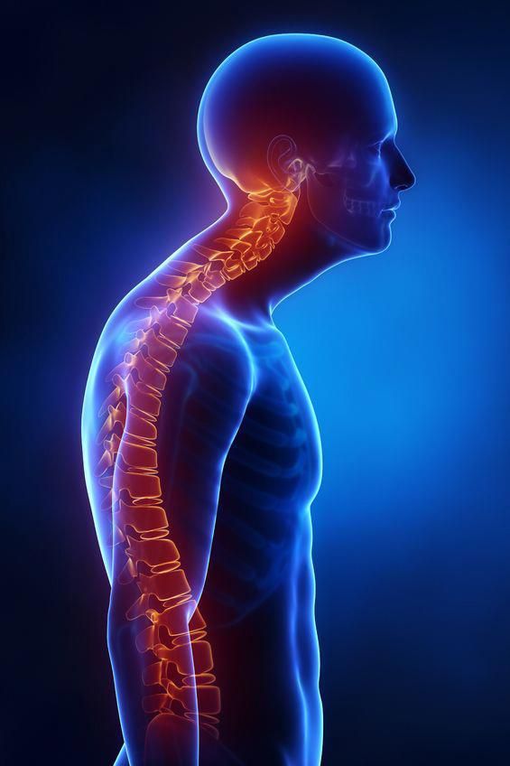
Kyphosis, often perceived as solely a spinal issue, significantly impacts overall posture and movement patterns. The compensatory mechanisms adopted by the body to maintain balance in the presence of kyphosis can place abnormal stresses on various joints, particularly the hips, leading to pain, dysfunction, and even injury.
Etiology
The primary cause is the altered biomechanics due to kyphosis. This can lead to:
- Anterior Pelvic Tilt: To compensate for thoracolumbar kyphosis, the pelvis often tilts anteriorly, increasing lumbar lordosis and hip flexor tightness.
- Altered Gait Mechanics: Changes in weight distribution and stride can put excessive stress on hip joints and surrounding soft tissues.
- Increased Joint Compression: Abnormal forces across the hip joint can lead to degenerative changes over time and cartilage wear.
- Muscle Imbalances: Weakness in hip extensors (glutes) and abdominal muscles, coupled with tightness in hip flexors and hamstrings, is common.
Clinical Features
Patients may present with:
- Visible forward rounding of the upper back.
- Fatigue.
- In severe cases, neurological symptoms (numbness, weakness) Especially hip internal rotation and hip extensor.
- Back pain may be present, but rarely is it significant enough to impact normal activity.
- Tight hamstrings (back thigh) muscles.
- Noticeable alterations in walking or standing posture.
Classification:
The deformity may be divided into three degrees according to its severity:
1. First degree
2. Second degree
3. Third degree
Progress of the deformity from first to third degree:
A bad habitual posture is the precipitating factor. Initially (first degree), if not corrected at this stage, it progresses to the second degree.
(a) The pectoral muscles become short, thereby restricting the chest expansion.
(b) Longitudinal back muscles, rhomboids and the middle trapezius are unduly stretched and weakened with loss of tone.
(c) Posterior ligaments are lengthened with corresponding shortening of the anterior structures. This gives rise to increased posterior laxity and a typical kyphotic deformity.
(d) During the adolescent stage of the growth period, wedging of the vertebral bodies may occur.
Investigation
Diagnosis involves a comprehensive assessment:
- Clinical Examination: Postural analysis, muscle strength testing ,range of motion assessment, , and palpation.
- Imaging (X-rays, MRI): To rule out other hip pathologies and assess the degree of spinal kyphosis. X-rays can show degenerative changes, while MRI can visualize soft tissue injuries.
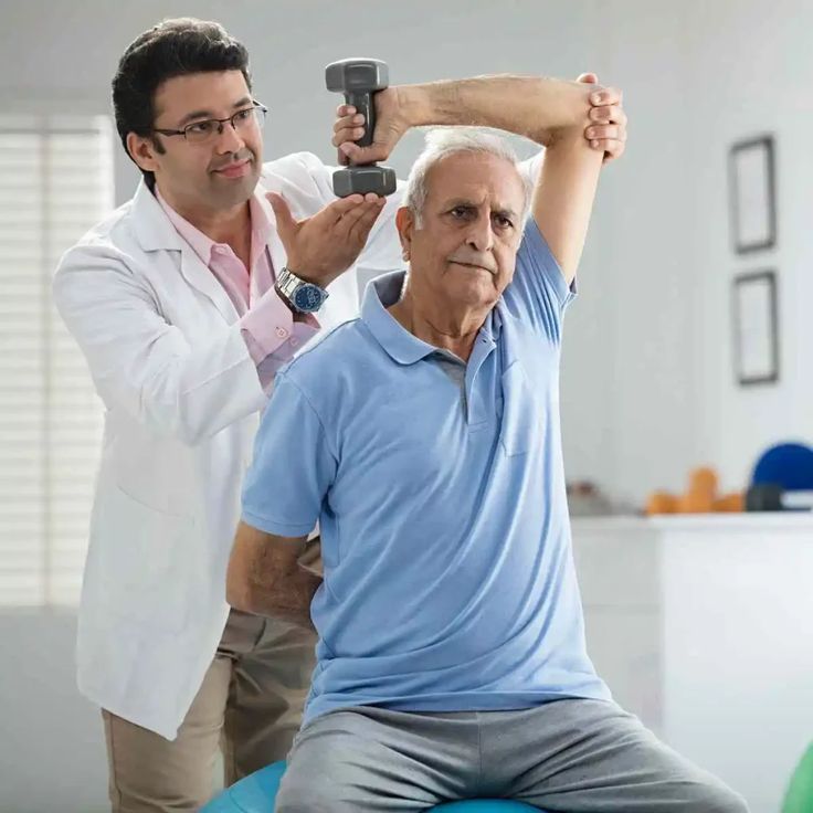
Physiotherapy Management
Physiotherapeutic management basically depends upon the stage of the condition and its ill effects.
First degree kyphosis:
It is postural and should be detected early.
1. Relaxation of the body, especially the upper back.
2. Repeated stretching sessions of shortened anterior structures by bracing the shoulders.
3. Maintaining posture of the head, neck and shoulder during activity or rest in optimal position should be trained and checked
4. Mobilization of the whole spine, particularly neck, scapulae and shoulders.
5. Diaphragmatic and costal breathing with emphasis on inspiration.
6. When precise mobility is attained, specific isometric and resistive exercises can be added.
7. Efficiency of a home management programme is vital for good results. Therefore, it needs to be emphasized and checked regularly.
8. The efficiency of home management in the form of watchful monitoring and correction is important.
Second and third degree kyphosis:
As wrong adaptation of the soft tissues is in the advanced stage, its active correction and maintenance are difficult.
The Milwaukee brace is prescribed with pads applied on the posterior uprights.
Exercises: With the brace on, the patient is encouraged to apply maximum pressure over these posterior pads. It stretches the shoulders, shoulder girdles and the kyphotic curve. Sustenance of this active stretching is very important.
It is difficult to achieve enough correction but it certainly helps in preventing further deterioration of the curve. Exercises to improve mobility and respiration reduce the overall impact of the deformity.
Diet and Nutrition in Kyphosis Management
Diet plays a vital role in supporting musculoskeletal recovery and posture correction. A balanced, nutrient-rich diet can aid bone health, muscle function, and inflammation control.
Recommended Dietary Tips:
- Include calcium and vitamin D-rich foods (like milk, yogurt, leafy greens, and sunlight exposure) to strengthen vertebrae.
- Add protein sources (egg, lentils, fish) to rebuild weakened back and core muscles.
- Consume anti-inflammatory foods such as turmeric, ginger, and omega-3–rich fish to reduce stiffness.
- Stay hydrated to maintain spinal disc elasticity and joint mobility.
- Avoid excessive processed sugar and junk food, as they can contribute to inflammation and muscle weakness.
Conclusion
An early kyphosis diagnosis leads to the best outcome. Most people who receive an early diagnosis don’t need surgery and can manage the condition with nonsurgical options. Kyphosis can get worse if left untreated. Addressing the underlying kyphosis is paramount for long-term hip health.
Q1. What is the main goal of physiotherapy in Kyphosis?
A1. To correct posture, stretch tight muscles, strengthen weak back muscles, and prevent deformity progression.
Q2. Which exercises are helpful in early-stage Kyphosis?
A2. Stretching of anterior muscles, spinal mobilization, breathing exercises, and postural correction training.
Q3. What device is used for advanced Kyphosis management?
A3. The Milwaukee brace is used to support correction and prevent further spinal curvature progression.