Introduction:
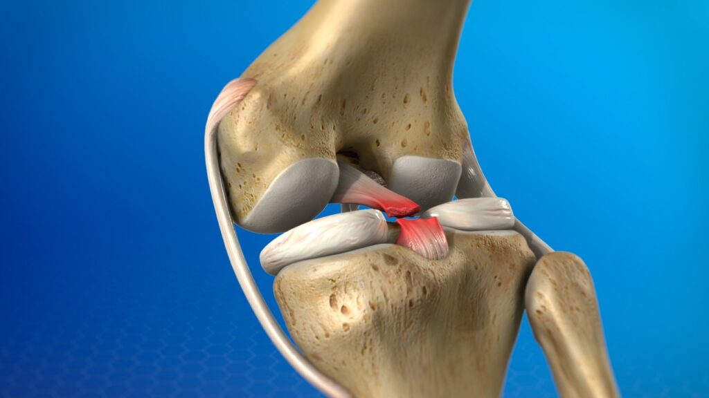
Posterior Cruciate Ligament Injuries occurs following a direct blow to the front of the knee. It may be associated with fracture of patella or even posterior dislocation of the hip.
Etiology:
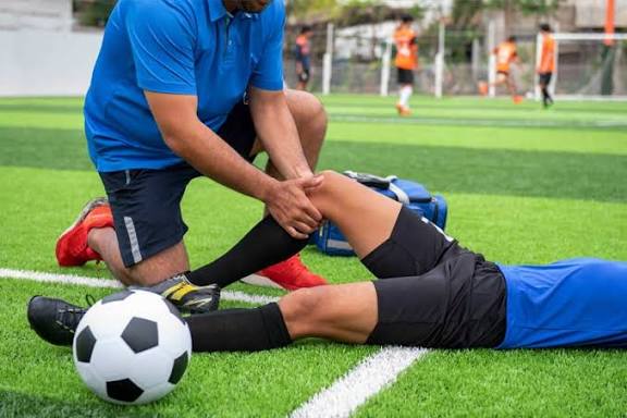
PCL injuries typically result from high- energy trauma. They are frequently seen in athletes involved in contact sports and individuals who have sustained road traffic accidents. Unlike Anterior Cruciate Ligament Injury(ACL)injuries which often occur with twisting motion, PCL tears usually result from direct blows to the front of knee.
Mechanism of Injury:
The most common mechanism of PCL injury is a direct blow to the front of the tibia with the knee bent. Hyperextension of the knee or a sudden, forceful push on the shin bone while the foot is planted can also lead to PCL tears.
Characteristic Feature:
Posterior Cruciate Ligament injuries present in different degrees according to the severity.
Grade 1: Limited damage with only microscopic tears in the ligament, mostly as the result of an overstretch. It is still able to function and stabilize the knee.
Grade 2: The ligament is partially torn. There is a feeling of instability.
Grade 3: Complete ligament rupture or tear . This type of injury is mostly accompanied by a sprain of the ACL and/or collateral ligaments.
Signs & Symptoms:
- Instability: Feeling of the knee “giving way”, particularly when downhill or descending stairs.
- Swelling: May develop rapidly after the injury.
- Pain: Often localized to the back of the knee, especially with going downstairs or kneeling.
- Posterior Sag sign: A key clinical sign where the tibia appears to sag backward when the knee is bent to 90 degrees.
- Difficulty bearing weight: Though some individuals maight be able to walk, it can be very painful.
Physiotherapy management:
Acute stage of injury:
The main aim of physiotherapy in the acute stage is reduction of inflammation and pain.
1. Cry therapy – Ice packs for 15 min every hour.
2. Modalities like diapulse, interferential therapy ,ultrasound therapy and TENS can be used.
3. Compressive or pressure bandage with limb in elevation.
4. Protection of knee from the position of stress.
5. Early initiation of isometrics for the quadriceps. They should be done at least for 5 min every hour. Prevention of quadriceps wasting and reflex inhibition is important.
6. If cast is not advised, a small range of self-assisted relaxed gentle knee swinging could be initiated.
Subacute stage of injury:
1. Initiate mobilization with the patient sitting at the edge of the bed or table, injured limb fully supported by the sound limb.
2. The range and power of quadriceps and hamstrings should be recorded.
3. Passive exercise to be progressed to assisted active or active as early as possible. Self-assisted relaxed passive stretching of knee flexion is excellent for improving the ROM of flexion.
Diet And Nutrition
A balanced diet rich in protein, calcium, and anti-inflammatory foods such as fruits like mud apple, leafy greens, and omega-3–rich foods supports tissue repair, strengthens muscles, and promotes faster healing during physiotherapy sessions.
Conclusion:
In combined anteromedial and anterolateral instability, full range extension may cause anterior subluxation of the tibia. Therefore, full arc of extension should be avoided while exercising.
Q1. What causes a PCL injury?
A1. PCL injury usually results from a direct blow to the front of the knee or high-energy trauma, such as sports or accidents.
Q2. What are common symptoms of a PCL tear?
A2. Symptoms include pain at the back of the knee, swelling, instability, and a posterior sag sign when the knee is bent.
Q3. How does physiotherapy help in PCL injury recovery?
A3. Physiotherapy reduces pain and swelling, prevents muscle wasting, restores knee motion, and strengthens quadriceps and hamstrings.