Introduction
It extends from the sustentaculum tali of the calcaneus to the plantar surface of the navicular bone. The ligaments are so arranged that they facilitate certain movements while at the same time check excessive and harmful joint movements. Spring ligament complex injuries or calcaneonavicular ligament injuries refer to stretching sprains, tears, or ruptures of the plantar calcaneonavicular ligament complex and can affect one or more of the three portions.
Etiology
Spring ligament injuries can arise from both acute trauma and chronic overuse.
- Acute Trauma: Sudden twisting motions, or falls from a height, awkward landings, can cause an immediate tear or stretch.
- Biomechanical Abnormalities: Conditions such as excessive pronation or posterior tibial tendon dysfunction place increased stress on the spring ligament, making it more susceptible to injury.
- Overuse/Repetitive Stress: Chronic strain from activities like running, swimming, or playing, especially on uneven surfaces, can lead to breakdown and irritation of the ligament fibers.
Clinical Features
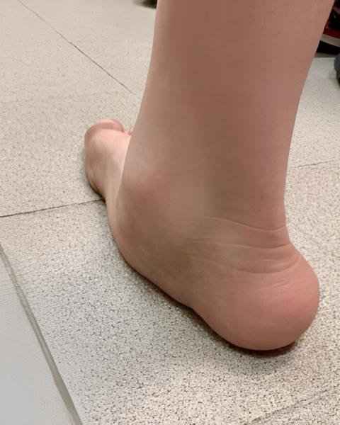
- Medial ankle and foot pain, especially during activity, and can lead to an acquired flatfoot deformity.
- Tenderness: There is often localized tenderness to palpation (touch) along the spring ligament, particularly between the talar head and navicular tuberosity.
- Pain that worsens with weight-bearing activities (walking, running, prolonged stand).
- In acute injuries, swelling and redness may be present around the medial ankle and foot.
- A sensation of instability or “giving way” in the foot.
Investigation
Diagnosis involves a comprehensive approach:
X-rays: X-rays are used to evaluate the alignment of bones and identify any fractures or dislocations that may be contributing to the symptoms.
Ultrasound: Can assess the spring ligament structure and associated tendon abnormalities.
MRI (Magnetic Resonance Imaging): Considered the gold standard for diagnosing spring ligament tears and evaluating co-existing issues like posterior tibial tendon pathology or mid-foot joint disease.
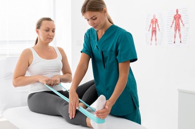
Physiotherapy Management
Conservative management is often the first line of treatment. Physiotherapy plays a crucial role:
Treatment for undisplaced ligament injury:
- Usually treated by a conservation approach of immobilization in a long leg cast for 6–8 weeks.
- Limb is maintained in elevation.
- Initiate vigorous, repetitive full ROM movements to the distal joints.
- Initiate early isometrics to the encased muscles.
- Diapulse, which can be given with the cast on, is ideal if available.
- NWB crutch walking is progressed to PWB first with crutches and with cane by 4–6 weeks.
- Kneecap or a crepe bandage is applied.
- Isometrics are intensified.
- Superficial thermotherapy, followed by rhythmic, relaxed, free, active knee mobilization, is emphasized.
- The patient is well educated and guided on the techniques of self assisted and self-stretched modes for knee flexion emphasizing sustaining and holding.
- Repetitive quadriceps exercises are performed against gravity.
- Active self-resistance exercises are important at this stage along with strong isometrics to the quadriceps.
- The best method of strengthening, increasing muscle power and endurance along with JROM is to teach and guide patients on self resistance and self-stretching modes of exercise.
- By 10–12 weeks, the patient should reach a stage of preinjury status. Treatment for displaced or completely torn ligament It may be a surgical emergency.
Dietary Support for Ligament Healing
- Protein-rich foods (like eggs, legumes, and lean meats) support collagen synthesis and muscle repair.
- Vitamin C (from oranges, amla, guava) aids in collagen formation, crucial for ligament healing.
- Calcium and Vitamin D (from dairy, leafy greens, and sunlight) strengthen bones and support the ligament-bone connection.
- Anti-inflammatory foods such as turmeric, nuts, and seeds help reduce swelling and pain naturally.
- Staying hydrated supports nutrient transport and tissue elasticity, enhancing recovery efficiency.
Conclusion
Spring ligament injuries, though uncommon, can lead to painful and potentially debilitating flatfoot deformities. These injuries often involve the superomedial (SM) band of the spring ligament complex and can be misdiagnosed as ankle sprains, leading to delayed treatment.