Introduction
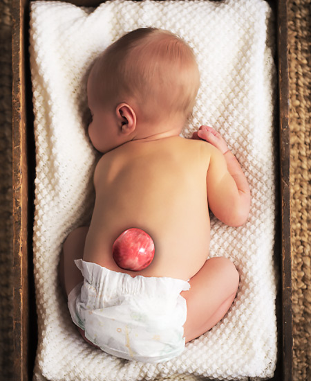
Spina Bifida is a birth defect that occurs when the spine and spinal cord don’t form properly. It falls under the umbrella of neural tube defects (NTDs), which are conditions affecting the brain, spinal cord or spine.
Definition
Spina bifida, a neural tube defect, is the result of the defective fusion of one or more posterior vertebral arches with resultant protrusion of the contents of the spinal canal.
Etiology
The exact cause of Spina Bifida is often multifactorial, involving a combination of genetic and environmental factors. Key contributing factors include:
- Folic Acid Deficiency: Insufficient folic acid intake during pregnancy and early pregnancy is a significant risk factor.
- Medications: Certain anti-seizure medications (e.g., valproic acid) can increase risk.
- Maternal Conditions: Conditions like uncontrolled diabetes, thyroid and obesity in the mother can elevate risk.
- Genetics: A family history of neural tube defects increases the risk.
Clinical Features
The signs and symptoms vary greatly depending on the type and severity of Spina Bifida. Common clinical features may include:
- Skin Ulceration: The presence of an overlying midline skin defect, such as haemangioma, lipoma or a tuft of hair.
- Motor Impairment: Weakness, paralysis or complication of paraplegia in the legs.
- Sensory Deficits: Reduced sensation below the level of the lesion.
- Bowel and Bladder Dysfunction: Incontinence, bladder and kidney infection is common.
- Orthopedic Issues: Clubfoot, patella dislocations, and scoliosis.
- Hydrocephalus: Accumulation of fluid in the brain, often requiring a shunt.
- Meningitis: Inflammation in meninges due to infection.
Classification
Spina Bifida is primarily classified into two main types
- Spina bifida occulta : there is merely a radiographic defect between the laminae.
2. Spina bifida cystica (manifesta): there is a developmental deficiency of laminae, spinous processes and the overlying muscles and skin.
(a) Meningocoele: In this variety, usually the spinal cord and its nerve roots are intact, only the covering membrane (meninges) projects as a dural sac.
(b) Myelomeningocoele: : Here, the spinal cord is exposed to the surface as a plaque or nervous tissue.
Investigation
Diagnosis typically involves:
- Prenatal Screening: Blood tests (alpha-fetoprotein, AFP), ultrasound, and amniocentesis.
- Postnatal Examination: Physical examination, X-rays, MRI, and CT scans to assess the extent of the defect.

Physiotherapy Management
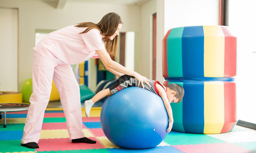
These children, therefore, do not require any specific treatment. Simple physiotherapeutic procedures can be effective to make them functionally independent.
Physiotherapeutic management includes the following:
- Prevention and management of deformities: Mild to moderate deformities may be treated conservatively by passive stretching and adequate splinting. Rigid deformities may need manipulation under general anaesthesia, followed by plasters and splints.
- Care of skin and joints : While using splints and braces and during passive stretching, protection of the skin from pressures and ulcerations is necessary. This is done by proper checking of the appliance and by frequent careful examination of the skin at pressure points.
- Management of bladder and bowel incontinence: Applying manual pressure above the symphysis pubis at regular intervals can be helpful in emptying the bladder in boys. In girls, it is difficult to obtain such control.
- Children who dribble constantly may develop ulcers at the napkin area, thus needing frequent examination of these areas and the necessary skin care.
- Infection of the urinary tract and failure of the kidney function can be avoided by surgical diversion of the urine to a receptacle.
- Education in ambulation, and self-care: Preparing a child for ambulation is the principal goal, and all the efforts to achieve this are initiated right from the beginning.
- Management of muscle paralysis: Depending upon the level of the lesion, priority should be given to: (a) Arm and shoulder girdle muscles. These should be developed for crutch walking and transfers. (b) Functionally important weight-bearing muscles such as hip abductors and extensors, knee extensors, ankle dorsi and plantar flexors should be strengthened to the maximum.
Diet and Nutrition in Spina Bifida
Proper nutrition plays a crucial role in prevention and rehabilitation of Spina Bifida:
- Folic Acid Intake: The most critical preventive factor. Women of childbearing age should ensure at least 400–800 mcg of folic acid daily through supplements or folate-rich foods such as spinach, citrus fruits, lentils, and fortified cereals.
- Balanced Diet: Include high-protein foods, fresh fruits, and vegetables to support tissue healing and muscle function.
- Hydration & Fiber: Adequate water and fiber help manage bowel function and prevent constipation, which is common in children with spinal defects.
- Calcium and Vitamin D: Essential for bone strength and to prevent orthopedic complications associated with limited mobility.
Conclusion
Spina Bifida presents unique challenges, but with early diagnosis, comprehensive medical care, and dedicated physiotherapy, individuals can achieve significant functional self-determination and lead activity of daily living. Understanding this state is the first step toward effective management and improved quality of life.
Q1. What is the main goal of physiotherapy in Spina Bifida?
A1. To promote mobility, independence, and prevent deformities through strengthening and functional training.
Q2. How does physiotherapy help prevent deformities in Spina Bifida?
A2. By using passive stretching, splinting, and corrective exercises to maintain joint alignment and flexibility.
Q3. What are key physiotherapy focuses in managing Spina Bifida?
A3. Skin care, bladder and bowel management, strengthening muscles, and training for ambulation and self-care.

